NB: This post is about three scans in Jan & Feb -- several months ago... It's taken me a long time to write about them. As for "now" -- I'm between scans therefore healthy :-) ... at least till the end of this month.
===
So, a picture is worth a thousand words? My own pictures seem to generate a thousand words of their own. Mostly incomprehensible words.
===
So, a picture is worth a thousand words? My own pictures seem to generate a thousand words of their own. Mostly incomprehensible words.
My last three scans generated a lot of excitement. What I mean is, worry. A new shadow on the brain, is it a new tumour? No it's not... Phew.
At the meeting with the cancer doc -- having found out that I am all clear -- I pick up a copy of the three scan reports. It's about time that I blog the actual results.
Scan one, MRI of brain:
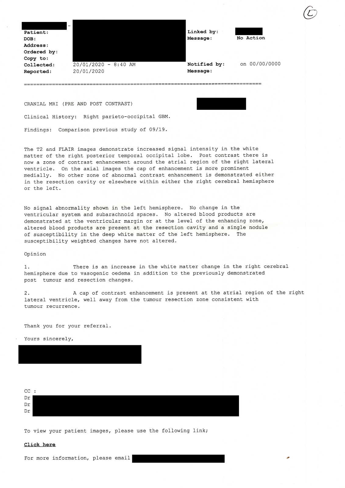
... "Increased signal intensity in the white matter of the right posterior temporal occipital lobe." I think that means, there are bad signs on the right of the brain.
... "Opinion: There is an increase in the white matter change... A cap of contrast enhancement... well away from the tumour resection zone consistent with tumour recurrence."
Which may mean that there is a potential new tumour but it's not where a new tumour is expected.
So, not a probability but a possibility. Enough to cause worry. Enough to get me lined up for another scan.
===
Scan two, just days later, PET of whole body:

Key points:
... "Head & Neck: There is only mild prominence of activity in the new enhancing lesion on MRI in the trigone of the right lateral ventricle." Which may mean that if it's a tumour it's not much of a tumour.
... "There is relatively reduced brain activity elsewhere..." Tough but fair "... associated with vasogenic oedema on MRI." Oh, I see, the reduced activity means less sign of an active new tumour.
... "Musculoskeletal: ... There is now less prominent degenerative activity around the left shoulder." My previous PET scan showed shoulder damage. I blamed RSI / OOS so changed my PC work posture. This PET shows that my changes worked. Amazing!
... "Interpretation: ... There is mildly increased FDG activity at the site of the likely tumour recurrence..." Which must relate to the "only mild prominence of activity" in the head & neck. I guess.
===
Scan three: MRI of brain, one month later

Key points:
... "Clinical history: Right Parietal GBM radiotherapy 2007." Well, 2017 actually. I'm sure that doesn't affect the conclusion.
... "The transverse dimension is identical ... with the previous imaging... No remote gliomatosis is identified." Nothing changed, nothing new. That's good news.
... "Conclusion: ... is thought to be related to treatment related phenomenon rather than progressive viable tumour." So there's the good news, damaged by the treatment :-)
===
There's a heap of medical jargon... some of it makes sense... knowing already what it means. Luckily the general thrust of it is interpreted for me.
===
Having held those pages for a couple of months I think it's time to file them with the other cancer-related paperwork. Except... I have already thrown out all earlier paperwork.
I know what cancers I have. I follow doctor's "suggestions". There's no need to read and understand all the fine medical details. Anyway, it can be depressing :-) And the doc holds all the originals. Let her be depressed...
The bulk of the paperwork was bills. Which are even less interesting than medical analyses. I did once add up the cost -- but it was paid, after that it is irrelevant.
I've finally "dealt with" these scan analyses. Interesting, yes. Worth keeping? no. Time to finally file them... out of sight, out of mind. Not immediately but later, in the bin.
Dr Nick Lethbridge / Consulting Dexitroboper
... Agamedes Consulting / Problems ? Solved
===
"No sense being pessimistic. It wouldn't work anyway." … per Ginger Meggs
===
"No sense being pessimistic. It wouldn't work anyway." … per Ginger Meggs
===
Dying for you to read my blog, at https: // notdotdeaddotyet .blogspot. com. au/ :-)
No comments:
Post a Comment